先日TVで、特殊能力を持つ人の脳内研究結果を見ました。
一度見た人の顔を記憶できるエスパーです。テロ対策に活躍していました。正面の写真を記憶するだけで、横顔だけでも認識できていました。その脳内では新しく記憶する度に海馬が増えているということでした。
そんな脳、私も欲しい!!!ところですが、開発して得られるものではないようでした。
脳って不思議。
BRAINの文字の引き寄せられた今日のVOAニュース。
医学にまつわる用語って、宇宙に関する用語と同じく、普段使わないので初めましてが多いです。
下記を頭に入れて、さぁ今日もLISTEN!!!
magnetic resonance imaging(MRI)
electrode(電極)
radioactive material (放射性物質)
schizophrenia 【 skìtsəfríːniə】 (統合失調症)
今日のVOAニュース!!!
科学者たちは今、脳のイメージング方法に疑問を呈している
Scientists Now Question Brain Imaging Methods
December 13, 2020
脳スキャンは珍しい光景を見せてくれます:心とその謎を覗くことができます。それらは多くの質問への答えを約束します。どうやって痛みを感じるのか?どうやって顔を知るのか?どうやって体を動かすのか?
しかし、脳スキャンは本当にこれらの疑問に答えられるのでしょうか?現在、多くの科学者が、脳スキャン研究の価値とその結果が真実かどうかを再考しています。
脳スキャン研究はいくつかの点で批判されてきました。批判には、被験者の数が少なすぎたり、結果を誤って読み取っていたりすることが含まれています。
また、研究者たちは、すべての条件が同じであっても、人の脳スキャンの結果は日によって異なることを理解するようになってきました。今では、脳スキャンの所見には限界があることを認めています。ある研究者は、これらの限界を研究しています。他の人は、異なる方法で脳を研究しています。
今年初め、ノースカロライナ州デューク大学の研究者であるアンチェン・クノット氏と彼女のチームは、このテーマに関する最新の論文を発表しました。それは、一般的な脳スキャンプロジェクトの有用性に疑問を投げかけたものです。チームは過去10年間に行われた60件の研究--彼女自身の研究も含めて調べました。
脳が’光る’のを見る
今回の研究では、機能的磁気共鳴画像法(fMRI)と呼ばれる手法を用いています。fMRIは、大きな磁石を使って、人が新しい言葉を覚えるなどの活動をしたときに、酸素を含んだ血液がどこに行くのかを調べるものです。これにより、科学者は脳の活動を測定することができます。
研究者がこの技術を使い始めたのは1990年代の初めです。当時、この技術は人間の脳への窓を開くものと思われていました。
それ以前のイメージング方法では、頭蓋骨の上に置かれた小さな装置--電極と呼ばれる--を通して脳の活動を調べることが行われていました。
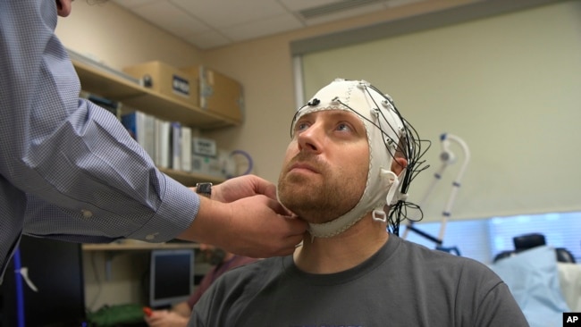 2019年11月20日の動画からのこの画像では、ザック・オルトは、マサチューセッツ州ベセスダにある国立衛生研究所の病院で、電極を使って脳の電気活動を追跡する脳波キャップを装着されています。(AP Photo/Federica Narancio)
血液中に放射性物質を注入する方法もありました。 それに比べて、fMRIの方が早くて簡単で 画像の質も高かったのです。
科学者たちはfMRIが脳の活動を 映し出すことについて 多くの論文を書き始めました。 報告者たちは多くの記事を書きました。 世間はもっと知りたいと思ったのです。
しかし、年月が経つにつれ、いくつかの研究結果には、厄介な兆候が現れ始めました。
ニューヨーク州アデルファイ大学の脳科学者であるダミアン・スタンレー氏は、「脳の画像を表示するのは非常に強力なものです」と語っています。しかし、このような強力なツールは、悪用や悪質な科学につながる可能性があるとスタンリー氏は言います。スタンレー氏はAP通信に、科学者の中には自分たちの研究が実際よりも重要であるかのように見せかけている人もいると言っています。
別のデューク大学の脳科学者アニータ・ディズニー氏はAP通信に「結局、我々はfMRIの流行に飛びつきすぎたのではないだろうか」と語ってくれました。言い換えれば、科学界がこの技術を支持し始めたのが、非常に早く、おそらく早すぎたということです。しかし今ではfMRIの問題は多くの科学者にとって懸念材料になっていると彼女は言います。
疑念が高まったことで、多くの研究室では、脳の研究にどの画像法を使うかについて慎重になってきました。そして、脳を研究するときには、見るべきものがたくさんあります。平均的な脳には約17万7000キロの神経線維があります。
イェール大学のジョイ・ハーシュ研究員は” 社会脳”を理解したいと考えています。 人が話したり、触れたり、目を合わせたりすると 脳内で何が起こるかを彼女は研究しています。ですから、彼女はfMRIは使いません。 一度に一人の人間にしか使えないので、その人間は大きなスキャナーの中で完全に静止していなければなりません。
その代わりに、ハーシュ氏は別の方法を使っています。頭部に取り付けられた光ファイバーケーブルにレーザー光を当てて血流を調べるのです。この方法では、スキャン中に被験者が自由に動くことができます。また、複数の人の間で実際に行われている社交性を研究することもできます。
デューク大学のアニータ・ディズニ氏ーもfMRIはあまり使いません。彼女は、fMRIは脳化学を研究するのに十分な精度を持っていないと説明しています。
しかし,誰もがfMRIから遠ざかっているわけではありません。
手術の前に患者の脳の地図を作るためにfMRIを利用している医師もいます。そして、fMRIは統合失調症やアルツハイマー病などの病気の研究にも有用であることが証明されています。
脳を研究する最新の方法
さて、新しいイメージング方法があります。それはオプトジェネティクスと呼ばれるものです。この方法は、光を使ってニューロンを活性化させます。脳科学の最新の研究方法かもしれません。しかし、科学者の中には、これをツールとして使うかどうかを知るには時期尚早だと言う人もいます。
ディズニー氏は、新しい方法が出てきたときには「その限界を理解するという基本的な作業を人にやってもらうのは、実際には本当に難しいことです。 」と指摘しています。
************
Scientists Now Question Brain Imaging Methods
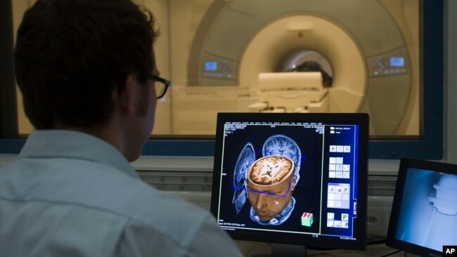 Sebastian Rieger, physicist MRI, makes a demonstration of the new 3 Tesla MRI (Magnetic Resonance Imaging) at the Brain and Behaviour Laboratory in Geneva, Switzerland, Dec. 8, 2008. (AP Photo/KEYSTONE/Salvatore Di Nolfi)
Brain scans offer us a rare sight: A look into the mind and its mysteries. They promise answers to many questions. How do we feel pain? How do we know a face? How do we move our body?
But can brain scans really answer these questions? Many scientists are now rethinking the value of brain scan research and whether its findings are true.
Brain scan studies have been criticized for several things. Criticisms include using too few subjects and incorrectly reading results.
Researchers have also come to understand that a person’s brain scan results can be different from day to day, even when all the conditions stay the same. Now they admit that brain scan findings are limited. Some are studying these limitations. Others are using different methods to study the brain.
Earlier this year, Annchen Knodt, a researcher at Duke University in North Carolina, and her team published the latest paper on this topic. It questioned the usefulness of common brain scan projects. The team looked at 60 studies of the past 10 years -- including Knodt’s own studies.
Watching brains ‘light up’
The research being re-examined uses a technique called functional magnetic resonance imaging, or fMRI. Using large magnets, fMRI scans find where oxygenated blood goes when someone does an activity, such as learning new words. This gives scientists a way to measure brain activity.
Researchers first began using this technology in the early 1990s. At the time, it seemed to open a window into the human brain.
Earlier imaging methods involved examining brain activity through small devices -- called electrodes -- placed on the skull.
Another technique involved injecting radioactive material into the blood. By comparison, fMRI seemed faster and easier And the images were of higher quality.
Scientists started writing many papers about fMRI’s ability to show brain activity. Reporters wrote many stories about these studies. The public wanted more.
But as years passed, troubling signs about some of the findings began to appear.
“It is a very powerful thing to show a picture of the brain,” said Damian Stanley, a brain scientist at Adelphi University in New York State. However, such a powerful tool, he said, can lead to abuse and bad science. Stanley told the Associated Press that some scientists made their research seem more important than it actually was.
Another Duke brain scientist, Anita Disney, told the AP, “In the end, we probably jumped on the fMRI bandwagon a little too fast.” In other words, the scientific community began supporting the technique very quickly, perhaps too quickly. But now, she said, the problems with fMRI are a concern to many scientists.
With doubts growing, many laboratories have become more careful about which imaging methods are used in brain research. And when studying the brain, there is a lot to look at. The average brain has about 177,000 kilometers of nerve fibers.
Yale University researcher Joy Hirsch wants to understand “the social brain.” She studies what happens in the brain when people talk, touch, or make eye contact with each other. So, she does not use fMRI. It can only be used on one person at a time and that person must stay perfectly still inside a large scanner.
Instead, Hirsch uses another method. Laser lights hit a fiber optic cable attached to a person’s head and then finds blood flow. This method lets her subjects move freely during scanning. It also permits her to study real-life socializing among several people.
Duke’s Anita Disney also does not use fMRI very often. She explained that it is not accurate enough to study brain chemistry.
But not everyone is walking away from fMRI.
Some doctors depend on the method to map a patient’s brain before an operation. And fMRI has proven useful for studying diseases such as schizophrenia and Alzheimer’s.
Latest method for studying the brain
Now, there is a new imaging method. It is called optogenetics. This method uses light to activate neurons. It may be brain science’s latest method of studying the brain. But some scientists say it is too early to know whether they will use it as a tool.
Disney noted that when a new method comes out “it is actually really difficult to get people to do the basic work of understanding its limitations.”
________________________________________________________________
Words in This Story
scan – n. the act or process of using a special machine to see the inside of something (such as a part of the body) scan – v. to examine especially systematically with a sensing device (as a photometer or a beam of radiation)
subject – n. a person or thing that is being dealt with in a particular way
jump on a bandwagon - idiom to join an activity that has become very popular or to change your opinion to one that has become very popular so that you can share in its success
neuroscientist – n. one who studies the scientific study of nerves and especially of how nerves affect learning and behavior
nerve fiber – n. any of the processes (such as axons or dendrites) of a neuron
fiber optic cable – n. technical : the use of thin threads of glass or plastic to carry very large amounts of information in the form of light signals
activate – v. to cause (a device) to start working
basic – adj. the simplest and most important parts of something (such as a subject of study)